Introduction
Subcutaneous nodules are palpable lumps located beneath the skin. They may be benign, inflammatory, infectious, metabolic, or malignant. A careful history, physical examination, imaging, and biopsy (when indicated) help determine the underlying cause.
SUBCUTANEOUS NODULES
Differential Diagnosis
1. Lipoma
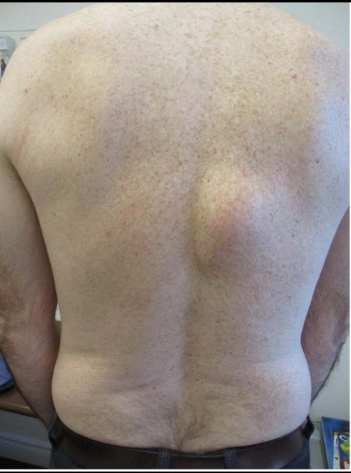
- Soft, mobile, painless fatty tumor.
- Most common benign subcutaneous nodule.
2. Epidermoid (Sebaceous) Cyst
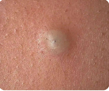
- Slow-growing, firm nodule with a central punctum.
- May become inflamed or infected.
3. Rheumatoid Nodules
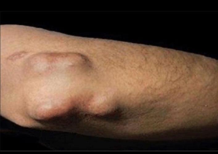
- Firm, non-tender nodules over pressure points.
- Common in patients with rheumatoid arthritis.
4. Erythema Nodosum
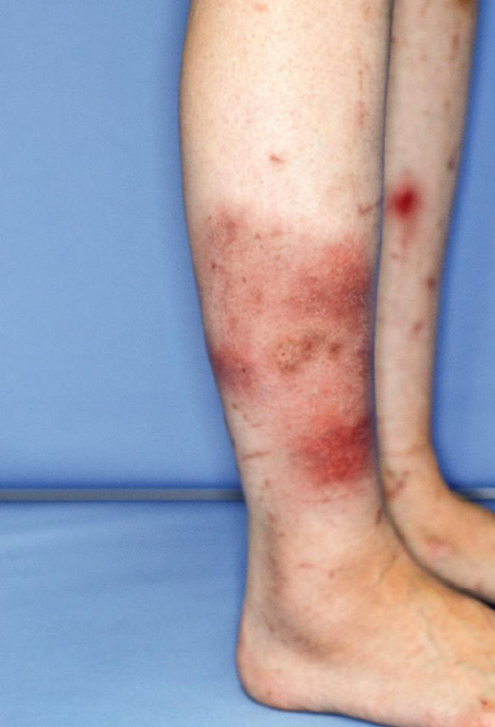
- Painful, red nodules, usually on the shins.
- Associated with infections, medications, autoimmune diseases, and pregnancy.
5. Gouty Tophi
- Hard nodules caused by uric acid crystal deposition.
- Common around joints, ears, fingers, and toes.
6. Dermatofibroma
- Small, firm, brownish skin nodule.
- Characteristic “dimple sign” on lateral compression.
7. Calcinosis Cutis
- Calcium deposits beneath the skin.
- Seen in connective tissue disorders and chronic kidney disease.
8. Abscess
- Painful, warm, fluctuant swelling containing pus.
- Often associated with fever and local redness.
9. Enlarged Lymph Nodes
- Mobile or fixed nodules depending on the cause.
- May result from infection, lymphoma, or metastatic cancer.
10. Soft Tissue Tumors
- Includes benign and malignant tumors (e.g., sarcoma).
- Rapid growth, pain, or fixation raises suspicion for malignancy.
Diagnostic Approach
- Detailed history (duration, pain, trauma, fever, weight loss)
- Physical examination
- Complete blood count (CBC), ESR, CRP
- Uric acid and autoimmune tests when indicated
- Ultrasound for cystic vs. solid lesions
- MRI for deep or suspicious masses
- Fine-needle aspiration cytology (FNAC) or biopsy for definitive diagnosis
Red Flag Signs
- Rapidly enlarging nodule
- Size greater than 5 cm
- Persistent pain
- Ulceration or bleeding
- Fixed to underlying tissue
- Associated unexplained weight loss or fever
Treatment
Treatment depends on the underlying diagnosis:
- Lipoma: Observation or surgical excision
- Epidermoid cyst: Excision if symptomatic
- Abscess: Incision and drainage with antibiotics if needed
- Erythema nodosum: Treat the underlying cause and provide anti-inflammatory therapy
- Rheumatoid nodules: Optimize rheumatoid arthritis treatment
- Gouty tophi: Urate-lowering therapy
- Malignant tumors: Multidisciplinary oncologic management
Frequently Asked Questions (FAQs)
1. Are all subcutaneous nodules cancerous?
No. Most subcutaneous nodules are benign, such as lipomas and epidermoid cysts.
2. When should I see a doctor?
If the nodule grows rapidly, becomes painful, bleeds, or persists for several weeks.
3. Can a lipoma disappear on its own?
No. Lipomas usually remain stable and require removal only if symptomatic or cosmetically bothersome.
4. Which imaging test is best?
Ultrasound is often the first investigation. MRI is preferred for deep or suspicious lesions.
5. Is a biopsy always necessary?
No. A biopsy is recommended when the diagnosis is uncertain or malignancy is suspected.
Conclusion
Subcutaneous nodules have a broad range of causes, from harmless lipomas to serious soft tissue malignancies. A systematic clinical evaluation combined with appropriate imaging and histopathological examination is essential for an accurate diagnosis and timely treatment. Early assessment of suspicious nodules improves outcomes and helps prevent complications.